Learn about rare inherited diseases that affect the retina and cause vision loss.
What is inherited retinal dystrophy (IRD)?
You might be wondering what IRD is, where it comes from, and what having an IRD will mean for you. This website is here to guide and support you.
So, what is IRD?
IRD is used to describe a collection of rare eye problems that are passed down from parents to children in the genes. IRDs are sometimes also called inherited retinal diseases or inherited retinal degenerations.1
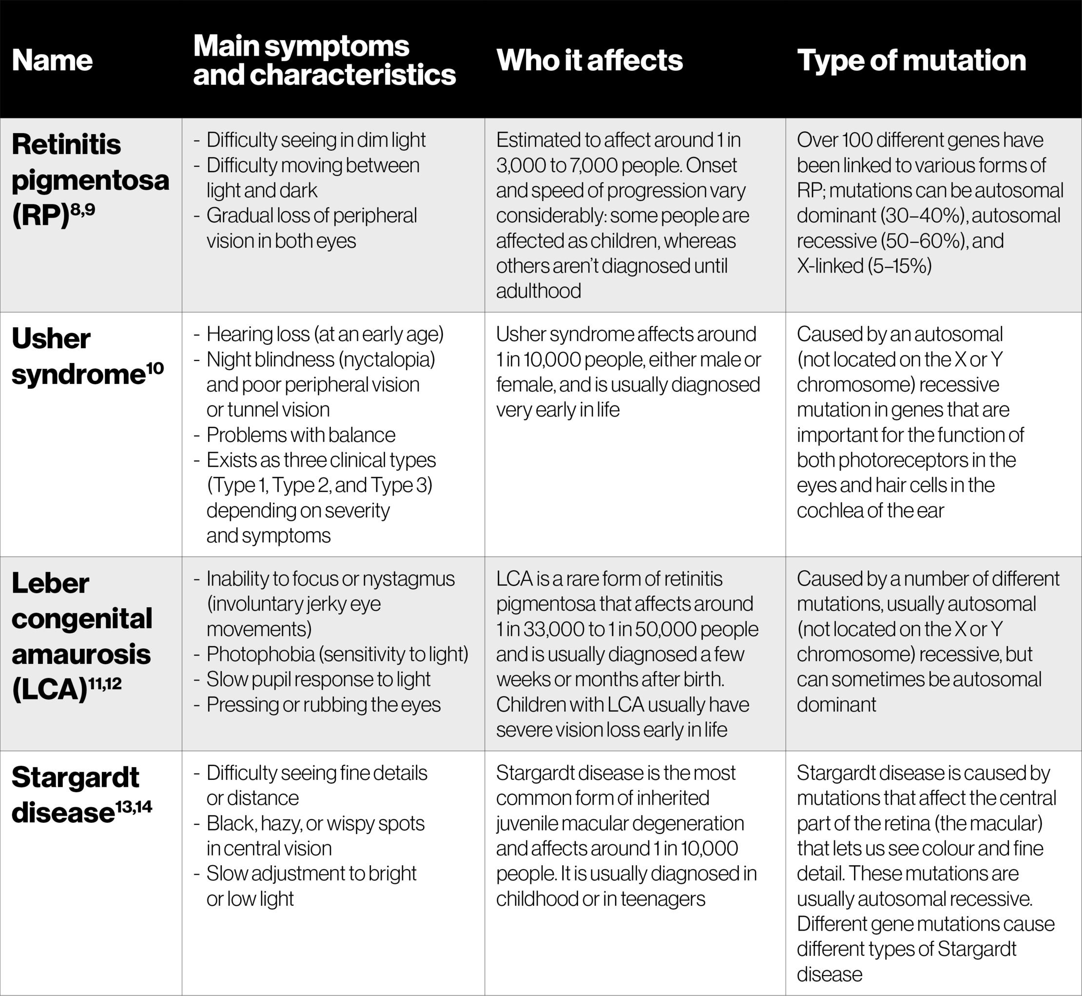
There are lots of different types of IRD. Some names of the more common ones are retinitis pigmentosa (RP), Usher syndrome, Leber congenital amaurosis, and Stargardt disease, all of which you can find more information on here
People with IRD have a mutation in one of the genes responsible for the function of the light-sensitive (photoreceptor) cells in the retina at the back of the eye. In IRD, these cells begin to stop working, work less effectively, or die, leading to a gradual loss of vision and blindness.1
It is important to remember that you are not alone in this. There are people of all ages living with an IRD, and it is the main cause of vision loss in people aged 15 to 45 years as well as a common cause of vision problems in children. Although rare, it has been estimated that IRD affects more than 2 million people worldwide (about 1 in every 2000).2
How different types of IRD affect the vision, symptoms and progression of these diseases are all different. This is because there are so many different types of IRD.
A quick guide to the retina
Retina: the back surface of the eye made up of two layers, full of cells that detect light (photoreceptor cells) and send signals to the brain to create images. Contains two types of photoreceptor cells: rods and cones1
Macula: the most light-sensitive part of the retina, densely packed with photoreceptor cells, a central spot that is responsible for colour vision
Rod cells: mostly found around the edge of the retina, these cells are the ones that work in dim light and that help us see things that aren’t straight ahead1
Cone cells: concentrated in the centre of the retina and mostly involved in seeing colour and give us detailed vision1
Gene mutations and IRD
Learning about IRD in more detail can equip you with a better understanding of your condition and help you come to terms with your diagnosis.
All forms of inherited retinal dystrophy are caused by mutations in genes. These genes are involved in the development and function of light-sensitive cells (photoreceptors) and other cells in the retina.1 More than 260 genes have been associated with IRD, and a genetic diagnosis can be found in about 60% of all cases of IRD.3,4
Some types of IRD (such as Stargardt disease) are mainly caused by one particular mutation.2 Others, like retinitis pigmentosa, can be caused by lots of different mutations but are still the same disease.2
In some cases the genetic mutation that causes an IRD isn’t yet known, but research is always being done to discover more. Genetic testing can confirm a genetic basis for your disease and may determine what mutation is responsible for your particular IRD and what your options are.
Common genetic terms
Medical language can feel very jargon heavy and full of words you may not understand. Here, are some of the most common words with quick definitions to make things easier.
- Autosomal: a mutation that is not on the X or Y chromosome
- X-linked: a mutation that is on the X chromosome
- Recessive: means that you need two copies of the mutated gene for the disease to cause symptoms
- Dominant: where only one copy of the mutation is needed for a person to be affected
- Carrier: Someone who has a copy of a recessive mutation but is not affected by the disease associated with it. A carrier can pass the mutated gene on to their children
Inheriting IRD
You may wonder how you came to have an IRD. All IRDs are inherited, which means you get the genes that cause them from your parents. Different types of IRD have different inheritance patterns depending on the type of mutation and the gene(s) affected. Below are the different ways that mutations that cause IRD can be inherited.

Common types of IRD
There are a lot of different conditions that together are called IRD. Many are named after the person who first described them. They can be caused by a mutation in one of over 260 genes that have been linked to these conditions.3
Types of IRD can be described as syndromic or non-syndromic. Syndromic IRDs are ones in which the eye problems are associated with other symptoms (such as hearing loss in Usher syndrome) as part of a systemic disease, whereas non-syndromic IRDs don’t have these systemic symptoms.6
Some of the more common ones are described below. As there are many others, you may have a type that isn’t included here. Other types of IRD can include: Bardet–Biedl syndrome, Joubert syndrome and primary ciliary dyskensia (among others).7
Disease progression
Knowing the facts can put you at ease
The thought of your condition progressing can be unsettling, but knowing the facts and what to expect can put you at ease and help you plan for the future.
How quickly an IRD is likely to progress and what symptoms you will develop can depend a lot on what form you have and the particular mutation. Even the same IRD can progress differently in different people. Many conditions start with difficulty seeing in low light levels, before leading to further vision problems. Loss of vision can be slow and gradual or can happen more suddenly. The different examples of visual impairment below help you to understand how the disease can develop.

Tunnel vision or loss of peripheral vision

Spots in the centre of vision

Blur in the centre of vision
